What is strabismus surgery?
Strabismus surgery strengthens or weakens eye muscles, which changes the alignment of the eyes relative to each other.
What kind of anesthesia is used for strabismus surgery?
The type of anesthesia depends on age/health and patient preference. Most children undergo general anesthesia. Adults typically have general anesthesia, conscious sedation or local anesthesia. The procedure is usually performed as an outpatient.
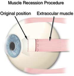
Fig. 1 In a recession procedure the muscle is cut from the surface of the eye and reattached further back from the front of the eye.
How does the surgeon approach the eye muscles?
The eye muscles attach to the sclera (wall of the eye). The muscles are covered by a thin layer of transparent tissue called the conjunctiva. The surgeon incises the conjunctiva to access the eye muscle(s), and uses a delicate hook to isolate the muscle. The eyelids are held open by a small instrument called a lid speculum. No skin incisions are made. The eyeball is NOT removed from the eye socket during strabismus surgery.
What is a recession?
A recession weakens function by altering the attachment site on the eyeball. A suture is placed through the muscle at the attachment site to the eye. The muscle is cut from the surface of the eye and reattached further back from the front of the eye [See figure 1].
What is a resection?
A resection strengthens function by reattaching a muscle to the eyeball at the original insertion site after a portion is removed. A suture is placed through the muscle at the intended new attachment site. The segment of muscle between the suture and the eyeball is removed and the shortened muscle is reattached to the eye [See figures 2 and 3].
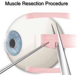
Fig. 2 In a resection procedure a segment of muscle is removed from the extraocular muscle. The shortened extraocular muscle is then reattached
What is an adjustable suture?
Strabismus surgery involves sewing the eye muscle to the wall of the eye after altering the insertion position and/or the length of the muscle. Standard strabismus surgery (no adjustable suture) utilizes a permanent knot. Adjustable suture technique utilizes a bow-knot or slip-knot (temporary knot) in an accessible position. After surgery the eye alignment can be altered by adjusting the temporary knot. The adjustment is typically done while awake and the operated eye numbed.
Are the eyes red after strabismus surgery?
It is normal for the white part of the eyes to be red after surgery. It may take several weeks/months for the redness to disappear. The eyes are usually scratchy and are sore upon movement. The soreness usually improves after a few days.
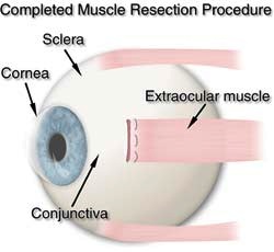
Fig. 3 After a resection the extraocular muscle has not changed position as in a recession. The only difference is the shorter length of the extraocular muscle.
Are patches or medicine used after surgery?
A patch is usually placed only if an adjustable suture is used. Some surgeons prefer an antibiotic or combination antibiotic/steroid drop or ointment after surgery.
information provided by http://www.aapos.org
What is it?
Strabismus, more commonly known as cross-eyed or wall-eyed, is a vision condition in which a person can not align both eyes simultaneously under normal conditions. One or both of the eyes may turn in, out, up or down. An eye turn may be constant (when the eye turns all of the time) or intermittent (turning only some of the time, such as, under stressful conditions or when ill). Whether constant or intermittent, strabismus always requires appropriate evaluation and treatment. Children do not outgrow strabismus!